
Product description
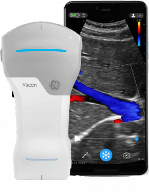
Vscan Air is a battery-operated general-purpose diagnostic ultrasound imaging system for use by qualified and trained healthcare professionals or practitioners. It enables ultrasound imaging guidance, visualization and measurement of anatomical structures and fluid. Vscan Air consists of a dual-headed probe, which integrates both curved and linear array transducers, and an app that can be installed on Android™ or iOS® mobile devices. Its pocket-sized portability and simplified user interface enables integration into training sessions and examinations in professional healthcare facilities (ex. hospital, clinic, medical office, home environment, road/air ambulance and in other environments described in the product user manual). The information can be used for basic/focused assessments and adjunctively with other medical data for clinical diagnosis purposes during routine, periodic follow-up, and triage assessments for adult, pediatric and neonatal patients. Vscan Air can also be useful for interventional guidance. Vscan Air customers have access to the Vscan web portal, including online access to product and product usage information for selected clinical scenarios.
- 128 physical channel beamforming
- Black-and-white mode for displaying anatomy in real-time
- Color-coded overlay for real-time blood flow imaging
- Harmonic imaging for increased signal-to-noise ratio and reduced artifacts from side lobes, grating lobes and reverberations, resulting in superior tissue definition and reduced speckle artifacts. With the greater penetration of lower ultrasound frequencies, high-quality harmonic imaging at greater depth can be performed.
- Selectable centre line marker
- Selectable focal zone marker
- Selectable TGC control with 6 depth-dependent gain controls
- Total scan time of 50 minutes with fully charged battery (with 80% black and white, 20% colour imaging)
- Any Qi-compliant wireless charger can be used to charge probe
- Recharge battery in 75 minutes from 10% to 90% battery capacity
- Dimensions: 131 x 64 x 31 mm
- Weight: 205 +/- 3 grams